
Translate this page into:
Rare case of Left Ventricular Hemangioma

Naga Sai Manjusha CH, MS Department of Cardiothoracic and Vascular Surgery, All India Institute of Medical Sciences New Delhi, 110049 India manjushasai89@rocketmail.com
This article was originally published by Thieme Medical and Scientific Publishers Pvt. Ltd. and was migrated to Scientific Scholar after the change of Publisher.
Abstract
Cardiac tumors are rare in occurrence. Among them, primary cardiac tumors that too hemangiomas are further rare. They usually are asymptomatic and are detected incidentally. Preoperative diagnosis may not correlate with histopathologic diagnosis. We report such a rare case of left ventricular hemangioma presented to us with vague chest discomfort, which preoperatively was suspected to be rhabdomyoma or fibroma.
Keywords
hemangioma
cardiac tumor

Introduction
Cardiac tumors per se are rare with frequency of approximately 0.001 to 0.030% at autopsy.1, 2 Though they can cause variety of symptoms including arrythmias, most of them are asymptomatic rendering themselves difficult to be diagnosed clinically, accounting to 5 to 10% of them being detected. Of those that are diagnosed, metastatic tumors are more common. Of all primary benign cardiac tumors, myxomas are most frequent while hemangiomas are rare. We report a case of patient with cardiac tumor, which initially suspected to be a myxoma or rhabdomyoma, turned out to be a hemangioma on histopathological analysis.
Case Report
A 12-year-old male patient presented to us with symptoms of vague intermittent chest pain and easy fatiguability. Clinical examination was inconclusive apart from tachycardia. Preoperative chest X-ray was normal. Electrocardiogram (ECG) showed sinus tachycardia. On echocardiography, there is mobile globular mass of size 2 × 1.78 cm with smooth surface, attached to mid interventricular septum on left ventricle (LV) side with thin stalk—suspected to be myxoma or rhabdomyoma (Figs. 1 2 3 4). On cardiac magnetic resonance imaging (MRI), there is an oval mobile mass lesion of size 1.8 (anteroposterior [AP]) X 1.4 (craniocaudal plane [CC]) cm in LV, prolapsing into LV outflow tract (LVOT) during systole. The lesion is isointense with late gadolinium enhancement on phase-sensitive inversion recovery. There was no LVOT obstruction (Figs. 5 and 6). Surgical approach was through median sternotomy followed by aortic and bicaval cannulation. After giving cardioplegia and cross clamping the aorta, transverse aortotomy was done, and aortic leaflets were retracted. There was a polypoidal pedunculated mass attached to LV septum. Pedicle was divided and the mass was retrieved. Aortotomy was closed, cardiopulmonary bypass was weaned off. Immediate postoperative period was uneventful. On Histopathological Examination (HPE) analysis, there was lobular proliferation of capillary sized vessels with diluted feeding vascular channels. Area near stalk and septal myocardial component showed variably dilated thin-walled vascular channels lined by thin flattened endothelial cells. Intervening stroma showed variable areas of fibrosis and myxoid changes without any adipose tissue or calcification. There was no hyalinization, atypia or necrosis. Immunohistochemistry revealed CD 31 (cluster differentiation), CD 34 and ERG-positive blood vessels. Androgen receptor and calretinin are negative. Overall features are suggestive of benign vascular tumor hemangioma, mixed type (polypoid capillary type and cavernous intraseptal component) (Fig. 7). Postoperatively, the patient was symptom free, was discharged on postoperative day 5, and echocardiography examination at 6 months follow-up revealed no residual mass.
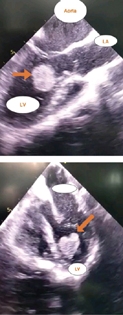
- Echocardiography—four-chamber view showing left ventricular mass. LA, left artery; LV, left ventricle.
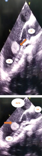
- Echocardiography—parasternal long axis view showing left ventricular mass. LV, left ventricle; LA, left artery.
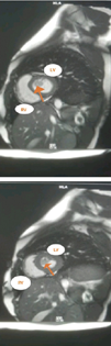
- Cardiac magnetic resonance imaging showing left ventricular mass. LA, left artery; LV, left ventricle; RV, right ventricle.
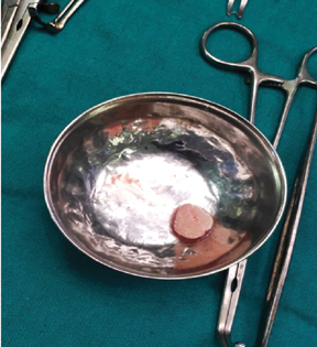
- Gross specimen of excised left ventricular hemangioma.
Discussion
Realdo Columbus first identified the presence of a heart tumor in 1559.3 Cardiac hemangiomas were first described by Uskoss in 1893,4 and the reported incidence is 2.8% of all cardiac tumors5 They can affect all age groups. They may arise from epicardium, myocardium, and pericardium. Intracavitary lesions include those arising from septum (interatrial and interventricular), from walls of chambers (both atria and ventricles) and over valves.6 The site of origin more commonly is LV (lateral wall) followed by right atrium (anterior wall) and least commonly involved is aortic valve. Hemangioma arising from interventricular septum, as was in the present case, is very rare. And those originating from septae are usually lethal due to more frequency of arrythmias. The symptoms depend on location, extent, and rarely histology of the tumor. Spectrum of presentation ranges from asymptomatic where the diagnosis is incidental to symptoms like exercise intolerance that is most common,5 followed by shortness of breath, chest discomfort, and rhythm disturbances including stokes Adam's syndrome, atrioventricular block, heart failure, pulmonary and systemic embolization causing cerebral infarcts,6 pseudoangina when tumor tissue compresses epicardial coronary arteries, angina pectoris, pericardial effusion and tamponade, LVOT or right ventricular outflow tract obstructive symptoms, failure to thrive. Rare presentation includes Kasabach–Merritt syndrome and cardiac failure. The mode of presentation in our patient was vague intermittent chest pain and easy fatiguability. In most cases, chest X-ray and ECG were unremarkable. Echocardiography is most useful tool in detecting cardiac tumors. A highly mobile LV tumor seen on echocardiography can give a superficial impression of a myxoma. Three-dimensional echocardiography can help to differentiate hemangioma from myxoma, through detection of densely packed echolucent spaces extending up to periphery of the lesion. In our patient, the lesion was suspected to be myxoma or rhabdomyoma. On MRI, compared with myxoma, the hemangioma has homogenous signal intensity after gadolinium injection and sequence of perfusion. Malignant tumors show infiltrative growth into the neighboring tissues, angiosarcoma has mosaic pattern in the T1 images, while hemangiomas are isointense.7 Angiography might show presence of tumor blush, characteristic of hemangioma due to pooling of contrast medium in sinusoids.7 Cardiac hemangiomas develop from endothelial cells mostly exophytic as pedunculated tumors, but intramural infiltrative growth is possible.7 Histologically, the tumor shows proliferation of blood vessels that may be either capillaries or large cavernous channels, with three subtypes: capillary, cavernous, and arteriovenous or combination of all the three types.5 Li et al concluded that the cavernous type (51.1%) was predominant subtype.5 Lack of mitotic activity, cellular pleomorphism, necrosis, and cellularity represent malignant transformation.6 In our case, the lesion is of mixed type and there was no atypia or necrosis showing benign nature of lesion.
Treatment
Treatment options include surgical removal with or without cardiopulmonary bypass depending on tumor location. Surgical removal is usually advocated as there is risk of intracavitary or valvular obstruction, peripheral embolization, and disturbances of rhythm or conduction. Approach may be through sternotomy or lateral thoracotomy. Care should be taken to prevent embolization, preserve myocardial function, and prevent conduction system damage, but complete resection of the tumor tissue it to be aimed at. Sometimes, these need extensive resection and reconstruction. Care should be taken for complete ligation of vascular channels, which if failed may lead to coronary cameral fistulae.8 There are a few case reports of either spontaneous regression or the tumor not growing in size, and being managed conservatively. But regular follow-up is must.6 Sometimes, the hemangiomas might be unresectable, which can be suspected through MRI and for such patients, steroid therapy and vincristine therapy may be tried.
Conclusion
This is a case of rare primary cardiac tumor, found on echocardiography in LV, without any characteristic clinical findings, that was suspected to be rhabdomyoma or fibroma. Instead, it was a hemangioma that is mobile in LV cavity, and surgical removal was done in view of high risk of embolization or LVOT obstruction. Echocardiography helps locating but MRI and fluorodeoxyglucose-positron emission tomography play role in determining nature of tumor and in suspecting malignancy and assessing resectability. Angiography helps in specifying diagnosis, ventricular function, and any fistulae. Surgical resection is the most important modality of treatment for primary benign cardiac tumors both curative and diagnostic. Newer modalities including vascular embolization will help us to increase the rate of detection and curability of these tumors. Regular long-term follow-up is essential as recurrences particularly in case of intracavitary benign tumors are known.
Conflict of Interest
None declared.
Funding None.
References
- Surgical pathology of primary cardiac and pericardial tumors. Eur J Cardiothorac Surg. 1997;12(5):730-737. , discussion 737–738
- [Google Scholar]
- Surgical treatment of cardiac neoplasms: 32-year experience. Thorac Cardiovasc Surg. 1990;38(02):176-182.
- [Google Scholar]
- Cardiac haemangioma with coronary-pulmonary artery fistula in one patient. Eur J Cardiothorac Surg. 2015;47(2):e77-e79.
- [Google Scholar]
- Case report: a rare case of pedunculated LV hemangioma evaluated by 3D echocardiography and compared with myxoma. Echocardiography. 2016;33(10):1608-1610.
- [Google Scholar]
- Cardiac hemangioma: a comprehensive analysis of 200 cases. Ann Thorac Surg. 2015;99(6):2246-2252.
- [Google Scholar]
- Non-invasive diagnosis of a pedunculated left ventricular hemangioma: tumor classification and evaluation of relevant literature. Clin Res Cardiol. 2007;96(4):227-231.
- [Google Scholar]


