Translate this page into:
A Rare Case of Unanticipated Difficult Intubation Due to a Vallecular Cyst
*Corresponding author: Chinmaya Nanda, Department of Cardiac Anaesthesia, Institute of Critical Care and Anaesthesiology, Medanta The Medicity, Gurugram, Haryana, India. nandachinu@gmail.com
-
Received: ,
Accepted: ,
How to cite this article: Nanda C. A Rare Case of Unanticipated Difficult Intubation Due to a Vallecular Cyst. J Card Crit Care TSS. 2024;8:252-3. doi: 10.25259/JCCC_14_2024
Dear Editor,
Vallecular cysts are extremely rare in adults and comprise approximately 10% of all laryngeal cysts.[1] They arise due to obstruction in the submucosal glands. Vallecular cysts are mostly asymptomatic. Sometimes, they present as dysphagia, voice change, stridor, or as a foreign body sensation. Newborns with a vallecular cyst may present with failure to thrive. Few case reports of difficult intubation due to a vallecular cyst have been described in the literature.[2,3]
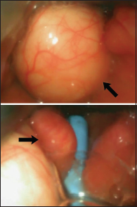
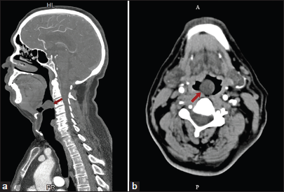
A 63-year-old male with a long-standing history of smoking and no other significant comorbidities was planned for a coronary artery bypass grafting. His airway assessment revealed a normal mouth opening with Mallampati grade II. His neck movements were not restricted. After standard induction with midazolam, fentanyl, etomidate, and vecuronium, a laryngoscopy was performed. A mass originating from the left side of the vallecula was observed obscuring the laryngeal inlet. After two attempts with the Macintosh Laryngoscope, a video-laryngoscope was utilized, and with the help of a bougie, the endotracheal tube was inserted after three more attempts [Video 1 and Figure 1]. The patient was hemodynamically stable and oxygen saturation remained stable throughout. The fiberoptic bronchoscope was also made available in the theater, but we succeeded in intubating the patient with the video-laryngoscope. The vallecular cyst was not excised in the same setting to avoid the risk of bleeding into the airway intra- and postoperatively. The patient was extubated the next day after discussing with the otolaryngologist. The rest of the recovery was uneventful. The patient was advised to follow-up with the otolaryngologist. A computed tomography scan of the head and neck was done later to assess the vallecular cyst [Figure 2].[4]

- Video-laryngoscopic view of the vallecular cyst and insertion of the bougie past the vallecular cyst (arrows).

- (a) Arrow showing cystic lesion in the vallecula on axial and (b) Sagittal images on computed tomography of head and neck.
Video 1:
Video 1:A cystic lesion is seen in the video-laryngoscope.An asymptomatic vallecular cyst can pose a serious challenge to the anesthesiologist due to unanticipated difficult intubation due to limited or non-visualization of the laryngeal inlet. The video-laryngoscope blade or the endotracheal tube can help push the cyst to visualize the laryngeal inlet. Bonfils intubation fiberscope or flexible fiberoptic bronchoscope can increase the success of securing the airway in such cases.
Ethical approval
The Institutional Review Board approval is not required.
Declaration of patient consent
The authors certify that they have obtained all appropriate patient consent.
Conflicts of interest
There are no conflicts of interest.
Use of artificial intelligence (AI)-assisted technology for manuscript preparation
The authors confirm that there was no use of artificial intelligence (AI)-assisted technology for assisting in the writing or editing of the manuscript and no images were manipulated using AI.
Video available online at
Financial support and sponsorship
Nil.
References
- Adult Vallecular Cyst: A Thirteen-year Experience. Otolaryngol Head Neck Surg. 2008;138:321-7.
- [CrossRef] [Google Scholar]
- Unanticipated Difficult Intubation as a Result of an Asymptomatic Vallecular Cyst. Anesthesiology. 1999;91:872-3.
- [CrossRef] [Google Scholar]
- Unexpected Difficult Intubation. Asymptomatic Epiglottic Cysts are a Cause of Upper Airway Obstruction during Anesthesia. Anesthesia. 1987;42:407-10.
- [CrossRef] [Google Scholar]


