
Translate this page into:
Severe Subglottic Tracheal Stenosis Dictates Intercontinental Transfer of a 2-Year-Old Child with Tracheostomy Tube In Situ
Manoj Kumar Sahu, MD, DNB Department of Cardiothoracic and Vascular Surgery CTVS office, 7th floor, CN Centre All India Institute of Medical Sciences New Delhi 110029 India drmanojsahu@gmail.com
This article was originally published by Thieme Medical and Scientific Publishers Private Ltd. and was migrated to Scientific Scholar after the change of Publisher.
Abstract
Atrioventricular septal defect (AVSD) is the most common congenital cardiac anomaly associated with Down’s syndrome. Children with AVSD develop pulmonary arterial hypertension and often require extensive therapy with pulmonary vasodilators in the postoperative period. The postoperative management is complicated by prolonged mechanical ventilation through endotracheal tube or tracheostomy tube. This artificial airway may trigger various airway complications including subglottic tracheal stenosis. The incidence and severity of subglottic tracheal stenosis is high in children with congenital syndromes. Inability to extubate or decannulate trachea and rapid respiratory compromise while attempting to do so directs toward a diagnosis of subglottic tracheal stenosis. The following case report discusses a 2–year-old boy from Nigeria who was discharged with tracheostomy tube in situ due to severe subglottic tracheal stenosis and surgical tracheoplasty could not be done at his present age. The implications of prolonged tracheostomy tube in situ and the hazards thereof during transfer of the child are being described.
Keywords
AV canal defect
Down’s syndrome
subglottic stenosis
tracheostomy

Introduction
Congenital1 cardiac repair of atrioventricular septal defect (AVSD) with pulmonary arterial hypertension in children with Down’s syndrome may be fraught with morbidities/complications postoperatively. Persistent pulmonary artery hypertension (PAH) may lead to prolonged invasive mechanical ventilation. Long duration endotracheal intubation or tracheostomy is a common cause of subglottic tracheal stenosis (SGTS).
Repeated manipulation of the endotracheal tube (ETT) or tracheostomy tube (TT) and breach in the tracheal mucosa with repeated tracheal suctioning may trigger SGTS. Prolonged airway instrumentation leads to mucosal ischemia which heals with formation of granulation tissue, therefore, constricting the airway.2
SGTS involves a partial or complete narrowing of the subglottic region of the airway (the area between vocal cords and cricoid cartilage) causing obstruction to free airflow and major respiratory complications.3 The reported incidence of postintubation and post-tracheostomy tracheal stenosis ranges from 10–19% to 8–65%, respectively.4 The use of high-volume low-pressure cuffed ETs/TTs and regulating the cuff pressure prevents development of SGTS.5 The clinical manifestations are in proportion to the degree of obstruction and the sufficiency of airflow. Mild stenosis may be misdiagnosed as laryngospasm, whereas more than 75% reduction in airway diameter present with severe respiratory distress6 and are classified as severe SGTS. This condition is diagnosed by radiological examination, indirect/direct laryngoscopy, or fiberoptic laryngo/bronchoscopy. Early recognition and management of SGTS leads to good long-term outcomes.2 The children with mild stenosis can be extubated, managed conservatively and sent home, whereas those with severe/critical stenosis need definitive treatment. Surgical tracheoplasty is the only definitive treatment option for high, moderate, or severe stenosis whereas endoscopic excision of stenotic segment, bronchoscopic dilatation, and tracheal stenting may be feasible in mild to moderate cases.7, 8 We in this report discuss about the difficulties with prolonged tracheostomy tube in a postoperative cardiac surgical child with critical SGTS and the anticipated problems during his transfer.
Case Report
A 2-year-old Nigerian boy, weighing 9 kg, known case of Down’s syndrome was diagnosed with complete balanced AVSD with increased pulmonary blood flow (Qp), patent ductus arteriosus (PDA), severe PAH, and normal biventricular function. The child developed bronchopneumonia preoperatively and was treated with intravenous antibiotics. Cardiac catheterization reported very high PAH 80/60 (34) mm Hg against aortic pressure of 90/64 (42) mm Hg, Qp/Qs = 2, and pulmonary vascular resistance (PVR) < 6 on oxygen. He successfully underwent an AVSD repair by 2-patch technique and PDA ligation. He was transferred to the intensive care unit (ICU) with a 5-mm uncuffed endotracheal tube. In the immediate postoperative period the patient developed low cardiac output syndrome (LCOS). A transthoracic echocardiography (TTE) revealed severe biventricular dysfunction, tricuspid regurgitation (gradient 67 mm Hg), and severe PAH (85 mm Hg) at a systemic pressure of 88/62 mm Hg. Pulmonary vasodilators like inhalational nitric oxide (iNO) 20 ppm (SV 300 NO, Siemens, Elema A B, Solna, Sweden), sildenafil (1.5 mg/kg/d) via nasogastric tube, and intravenous phenoxybenzamine (1 mg/kg/d) were started and the child was sedated and ventilated for next 48 hours. Although the ventricular function and LCOS improved on postoperative day (POD) 3, the pulmonary artery pressure remained equivalent to systemic pressure. Oral bosentan (4 mg/kg/d) was thus added on POD 4.
The child developed ventilation-associated pneumonia (VAP) with Acinetobacter spp. and was treated with imipenem and cefoperazone-sulbactum. Following resolution of pneumonia the child was weaned off iNO gradually. Repeated attempts at weaning from ventilatory support in subsequent days failed due to persistent severe PAH and residual hyperactive airways, often triggering acceleration in pulmonary artery pressures. A tracheostomy was performed on POD 9 and thereafter multiple weaning trials were attempted without success. Finally on POD 18, the child was separated from ventilator and put on T piece circuit on oxygen flow of 8 L/min. Until this time the child’s nutrition was maintained by enteral route, which he was tolerating well without much feed interruptions.
The patient developed a second episode of lower respiratory tract infection caused by multidrug resistant Pseudomonas which was treated with colistin. The child improved and the TT was downsized from 4.5 mm cuffed to 4 mm uncuffed one. A TTE showed good biventricular function with severe PAH. The child maintained peripheral oxyhemoglobin saturation (SpO2) of 90 to 95% on 1 L/min of oxygen supplementation by nasal cannula through tracheostomy tube and after a week he was weaned off oxygen completely (Fig. 1). On first tracheal decannulation trial, the child immediately started gasping for air and became cyanosed due to severe airway obstruction. The TT was reinserted instantly which provided sudden relief to the child and he started breathing normally through the TT. This event of decannulation made us certain that there was an obstruction above the tracheostomy level. A direct laryngoscopy done by otorhinolaryngologist confirmed critical SGTS (near 100% stenosis). A surgical tracheoplasty was advised, but only to be performed when the child attains an age of 6 to 7 years.
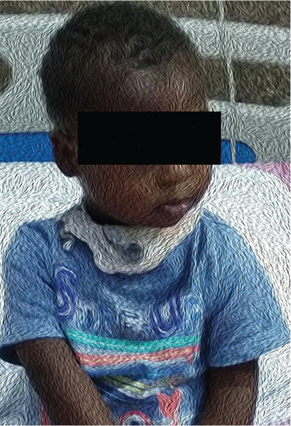
- The child with tracheostomy tube without oxygen supplementation in CTVS ward. CTVS, cardiothoracic and vascular surgery.
Not only did the transfer of this child with the TT in situ to a distant land by air seem hazardous but also practical difficulties in maintaining the TT by nonmedical personnel at home for years was a big concern for the authors. Extensive arrangements were made for his travel and the designated medical personnel with the airline were communicated about the child’s condition at two airports (as it was a stop by flight) to ensure his safe return. The mother was trained to take care of the TT on the way and at home. She was taught tracheal suctioning with foot operated portable suction machine, TT dressing changes with plain sterile gauze, changing the securing tie, to apply the nasal prong oxygen through the TT in case necessity arise and to follow-up with an otorhinolaryngologist in Nigeria earliest after their arrival. The foot suction machine, other accessories like spare TTs, appropriate size suction catheters, packed gauze, and so forth were supplied to the mother for the way. The airlines had arranged oxygen backup on the way. Finally the child flew to his home without any problem; he is now 1 year post cardiac surgery with TT and doing well (Fig. 2).
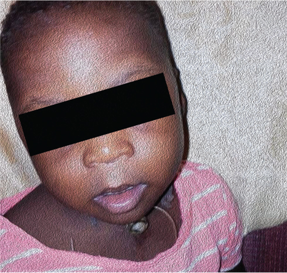
- The child with tracheostomy tube without oxygen at home at 8 months after surgery in Nigeria.
Discussion
Congenital heart disease is reported to occur in 40to 60% of patients with Down’s syndrome with complete AVSD being the most common defect.1 After surgical AVSD repair, postoperative morbidity remains common in this subset of population,9 PAH being one very important and challenging problems to treat. The presence of hyper-reactive airways in these children further aggravates the PAH, rendering weaning efforts from mechanical ventilation futile. In spite of anticipation and aggressive management of PAH in the early postoperative period, our patient had prolonged mechanical ventilation and developed lower respiratory tract infection.
Subglottic area is the narrowest part of the airway in children, which may be traumatized during endotracheal intubation (EI), or tracheostomy and prolonged endotracheal ventilation.2 Iatrogenic SGTS was mainly correlated to mucosal damage and ischemia induced by the rigid wall of the ETT or TT or cuff pressure and inflammatory response. Prolonged EI leads to chronic inflammation, fibrosis, and narrowing of the trachea.3
The potential risk factors for SGTS include the underlying disease requiring EI, the age and body weight, the duration and number of EIs, inadequate or no sedation during mechanical ventilation, and the occurrence of respiratory infections and hypotensive or hypoxic events during the period of EI and traumatic EI.10 In addition LCOS may cause tracheal mucosal hypoperfusion in pressure areas.
The young age, Down’s syndrome, narrower subglottic area, prolonged intubation, tracheostomy, mechanical ventilation, respiratory infections, and LCOS were identified as the possible risk factors in our patient.10 SGTS can be avoided by simple precautions like regulating the cuff pressure and avoiding recurrent, traumatic, and long-duration intubations and preventing oversized tubes, and so forth.5, 6
Classical presentations of SGTS is not seen in small children when ETT or TT is in situ. Clinically diagnosing an airway obstruction above tracheostomy level requires a high index of suspicion, especially when decannulation or extubation trail fails with typical presentation of sudden choking or gasping respiration and cyanosis. Careful supervised decannulation of TT or extubation should be attempted in these high-risk patients, so that sudden catastrophe is avoided in case of severe airway obstruction.
Implications of critical SGTS in a postoperative pediatric cardiac surgical patient are multifold—they need to be on long-term TT till final corrective surgery (tracheoplasty) is done. And the morbidities because of pronged TT include rapid acceleration of PAH, RV dysfuction (complicate the hemodynamics, may lead to LCOS), delayed weaning off invasive ventilation, higher chances of nosocomial infection including VAP, longer inotrope, vasodilator therapy, ICU stay, difficult to maintain nutrition, and overall turbulent postoperative convalescence. And in the longer run over the years the child’s overall development, particularly speech, will be affected. Inadvertent sudden catastrophes like blockade of the tube, sudden decannulation, repeated respiratory infections, increased secretions, and so forth may happen (particularly during travel/transport), so the child needs fulltime observation by the parents/caregiver. They should be taught about the TT care as described in case report. Overall managing SGTS in postoperative cardiac surgical patients is very heavy and challenging till the final surgical correction.
In conclusion, early and aggressive management of PAH in children with Down’s syndrome who undergo AVSD repair is prudent, to wean these patients from mechanical ventilation. SGTS should be ruled out in cases of extubation or decannulation failure. In children diagnosed with severe SGTS, it is possible to retain the TT in situ till the child is fit for surgery depending on age and nutrition status but requires continuous care and support by parents/ dedicated caregiver.
Ethical Approval
All procedures performed in this case were in accordance with the ethical standards of the institutional and/or national research committee and with the 1964 Helsinki declaration and its later amendments or comparable ethical standards.
Informed Consent
Written informed consent was taken from the parents.
Conflict of Interest
None.
Funding All the authors declare that they did not receive any funding from any source for this case study.
References
- Associated congenital anomalies among cases with Down syndrome. Eur J Med Genet. 2015;58(12):674-680.
- [Google Scholar]
- Postintubation tracheal stenosis: case report and review of current management. J Case Rep Med. 2013;2:1-3.
- [Google Scholar]
- A bronchoscopic approach to benign subglottic stenosis. SAGE Open Med Case Rep. 2017;5:X17713151.
- [Google Scholar]
- Complete subglottic tracheal stenosis managed with rigid bronchoscopy and T-tube placement. Lung India. 2016;33(6):661-663.
- [Google Scholar]
- Detection and management of tracheal stenosis following cuffed tube tracheostomy. Ann Thorac Surg. 1971;12(4):359-374.
- [Google Scholar]
- Benign tracheal stenosis a case report and up to date management. Ann Transl Med. 2016;4(22):451.
- [Google Scholar]
- Endoscopic management of subglottic stenosis. JAMA Otolaryngol Head Neck Surg. 2017;143(5):500-505.
- [Google Scholar]
- Operative and non-operative treatment of benign subglottic laryngotracheal stenosis. Eur J Cardiothorac Surg. 2004;26(4):818-822.
- [Google Scholar]
- Congenital heart surgery outcomes in Down syndrome: analysis of a national clinical database. Pediatrics. 2010;126(2):315-322.
- [Google Scholar]
- Complications and consequences of endotracheal intubation and tracheotomy. A prospective study of 150 critically ill adult patients. Am J Med. 1981;70(1):65-76.
- [Google Scholar]


